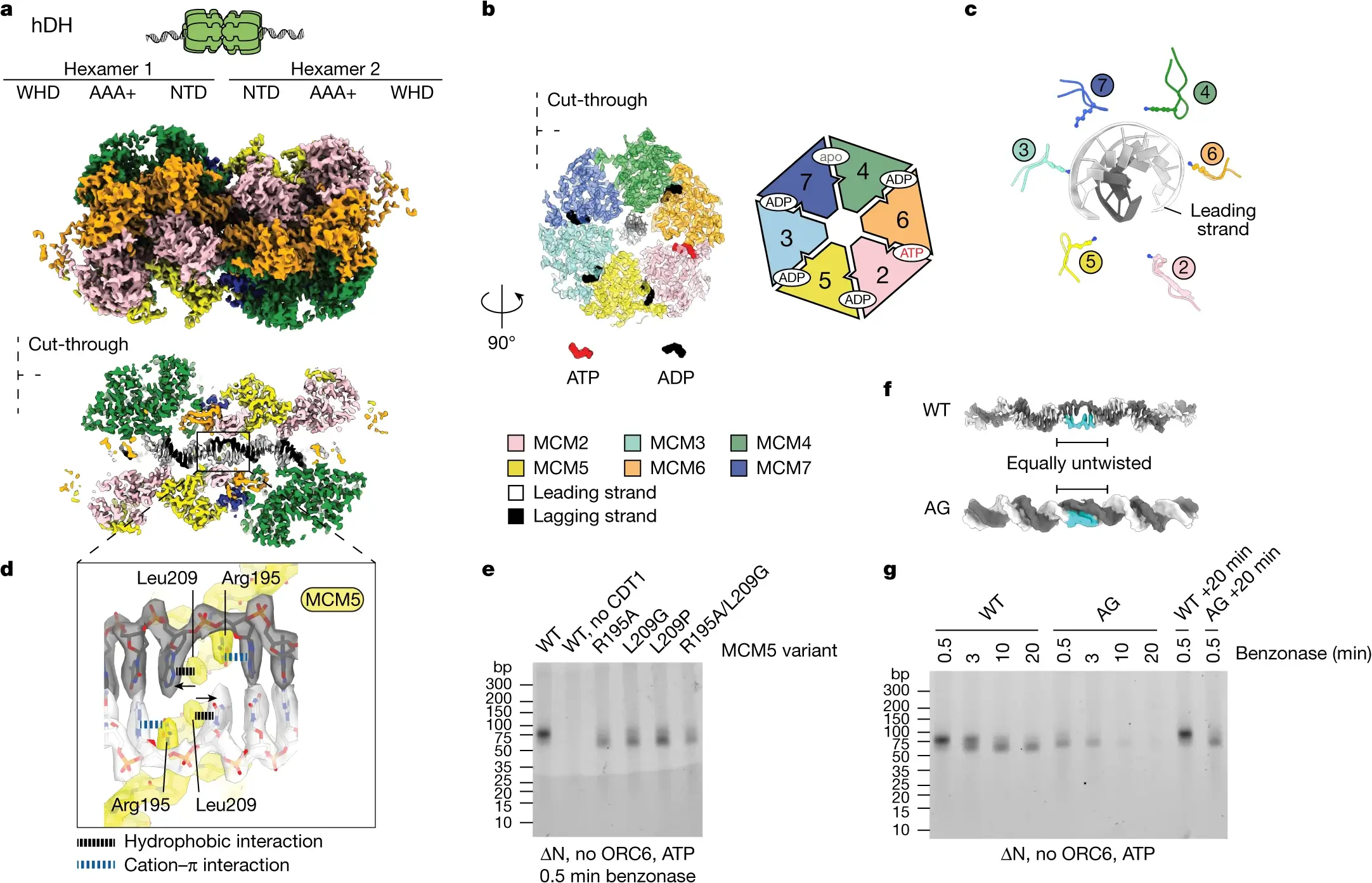
In a groundbreaking stride towards understanding the essential machinery of life, scientists are utilizing innovative imaging techniques to unravel the complex structures of human proteins. These microscopic powerhouses are pivotal to numerous biological processes, from cell signaling to immune responses. Despite their importance, visualizing the intricate architecture of proteins has remained a significant challenge—until now.
The Significance of Protein Structures
Proteins are the workhorses of cells, carrying out a myriad of functions necessary for life. Each protein’s function is intricately linked to its three-dimensional structure. Understanding these structures unlocks insights into how proteins interact with other molecules, how they contribute to health and disease, and how they can be targeted in drug design. However, the high complexity and minute size of proteins make them difficult to study.
Advanced Imaging: A New Frontier
Recent advances in imaging technology, including cryo-electron microscopy (cryo-EM) and X-ray crystallography, have revolutionized our ability to visualize proteins at atomic resolution. These techniques allow scientists to freeze proteins mid-action, capturing snapshots of their structures in near-native states. Cryo-EM, in particular, has been pivotal in revealing the structures of proteins that were previously deemed too complex to study.
Cryo-EM: Freezing Time to Capture Details
Cryo-EM involves rapidly freezing protein samples to preserve their natural shape, then bombarding them with high-energy electrons to produce detailed images. This technique enables researchers to construct three-dimensional models of proteins, revealing intricate details about their surfaces and active sites. Such insights are crucial for understanding how proteins work and how they can be manipulated for therapeutic purposes.
X-ray Crystallography: Unveiling Hidden Patterns
X-ray crystallography complements cryo-EM by offering another method to determine protein structures. It involves crystallizing proteins and then using X-ray beams to analyze how the crystals scatter the light. The resulting diffraction patterns provide a map of the protein’s atomic structure. This method has been instrumental in uncovering the structures of many critical proteins, such as enzymes and antibodies.
Implications for Medical Research
The ability to visualize proteins in detail has profound implications for medical research. With detailed structural information, scientists can design drugs that precisely target specific proteins involved in diseases. This precision reduces side effects and increases treatment efficacy. For example, understanding the structure of viral proteins can lead to the development of antiviral drugs that effectively disable viruses without harming the host cells.
Challenges and Future Directions
Despite the advances, challenges remain. Some proteins are difficult to crystallize or too dynamic to capture in a single conformation. However, ongoing developments in imaging technologies promise to overcome these hurdles. The integration of artificial intelligence in analyzing imaging data is enhancing our ability to interpret complex protein structures quickly and accurately, opening new avenues for research and drug discovery.
Bridging the Gap Between Structure and Function
Ultimately, these innovative imaging techniques are bridging the gap between understanding protein structures and their functions. As science advances, the detailed images of proteins not only deepen our understanding of biology but also empower researchers to tackle some of the most challenging diseases of our time.
In this rapidly evolving field, each imaging breakthrough brings us closer to a world where we can fully understand and harness the power of proteins, improving health outcomes and unlocking new scientific possibilities. Through these innovative techniques, the once hidden world of human proteins is now becoming a vivid reality, guiding the future of scientific discovery and medical innovation.


